What does skeletal muscle consist of? How does it contract?
Muscle anatomy can get quite complex… and that’s even without mentioning the physiology of muscle contraction! This article breaks skeletal muscle down into its smallest parts and examines the amazing processes that bring about all our movements…
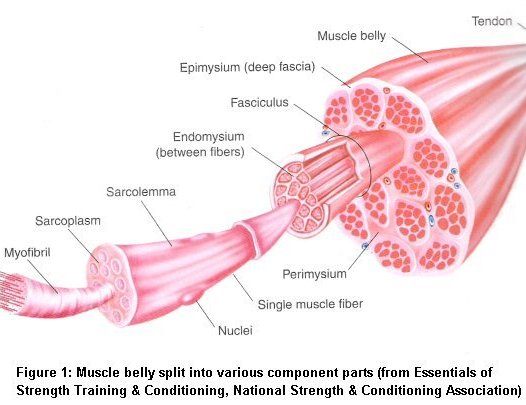
Every one of the body’s 430 skeletal muscles consists of muscle tissue, connective tissue, nerves and blood vessels. A fibrous fascia called the epimysium covers each muscle and tendon. Tendons connect the muscle belly to bone and they attach to the bone periosteum – more connective tissue that covers all bones. Contraction of the muscle belly pulls on the tendon and in turn, the bone it is attached to.
Limb muscles (such as the biceps brachii in the upper arm) have two attachments to bone. The proximal or origin is the attachment closest to the trunk. The distal or insertion is the attachment furthest from the trunk. Trunk muscles (such as the rectus abdominus in the stomach) also have two attachments – superior (closer to the head) and inferior (further from the head).
A closer look at muscle anatomy shows that each muscle belly is made up of muscle cells or fibres. Muscle fibres are grouped into bundles (of up to 150 fibres) called fasciculi. Each fasiculus or bundle is surrounded by connective tissue called perimysium. Fibres within each bundle are surrounded by more connective tissue called endomysium.
Each individual fibre consists of a membrane (sarcolemma) and can be further broken down into hundreds or even thousands of myofibrils. Myofibrils are surrounded by sarcoplasm and together they make up the contractile components of a muscle. See the diagram below:

Sarcoplasm contains glycogen, fat particles, enzymes and the mitochondria. The myofibrils it encases consist of two types of protien filaments or myofilaments. They are actin and myosin.
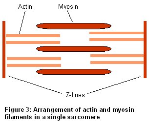
Myosin and actin filaments run in parallel to each other along the length of the muscle fibre. Myosin has tiny globular heads protruding from it at regular intervals. These are called cross bridges and play a pivotal role in muscle action. For more details, see the article on sliding filament theory.
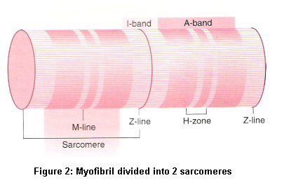
Each myofibril is organized into sections along its length. Each section is called a sarcomere and they are repeated right along the length of a muscle fibre. It’s similar to how a meter ruler is split into centimeters and millimeters. Just as the millimeter is the smallest function of a ruler, the sarcomere is the smallest contractile portion of a muscle fibre.
The sarcomere is often divided up into different zones to show how it behaves during muscle action. See the diagram below:

The Z-line seperates each sarcomere. The H-zone is the center of the sarcomere and the M-line is where adjacent myosin filaments anchor on to each other. On the diagram above the darker A-bands are where myosin filaments align and the lighter I-bands are where actin filaments align. When muscle contracts the H-zone and I-band both decrease as the z-lines are pulled towards each other. See the diagram below:

An examination of muscle anatomy wouldn’t be complete without taking a closer look at how muscle contracts…
Click here for the article on sliding filament theory
Sources
1) Baechle TR and Earle RW. (2000) Essentials of Strength Training and Conditioning: 2nd Edition. Champaign, IL: Human Kinetics
2) McArdle WD, Katch FI and Katch VL. (2000) Essentials of Exercise Physiology: 2nd Edition Philadelphia, PA: Lippincott Williams & Wilkins
3) Wilmore JH and Costill DL. (2005) Physiology of Sport and Exercise: 3rd Edition. Champaign, IL: Human Kinetics

Jacky has a degree in Sports Science and is a Certified Sports and Conditioning Coach. He has also worked with clients around the world as a personal trainer.
He has been fortunate enough to work with a wide range of people from very different ends of the fitness spectrum. Through promoting positive health changes with diet and exercise, he has helped patients recover from aging-related and other otherwise debilitating diseases.
He spends most of his time these days writing fitness-related content of some form or another. He still likes to work with people on a one-to-one basis – he just doesn’t get up at 5am to see clients anymore.